Alternative Cancer Secrets Ebooks
More healthy lungs x ray images. prescription cialis 5mg faq cialis for sale cheap xray viagra college essay help connecticut faustus essay reditabs uk cost buy cialis doctor online two viagras healthy man viagra from xmradio add accutane scars acheter One of the signs of copd that may show up on an healthy lungs xray x-ray are hyperinflated lungs. this means the lungs appear larger than normal. also, the diaphragm may look lower and flatter than usual, and the. The lungs should be well aerated without focal or diffuse areas of opacification. in this ap supine portable chest x-ray there is a sharply defined margin of the right lower lobe density (black.
Find Out Why Fat Cholesterol Salt Are Good For You A Fine Wordpress Com Site
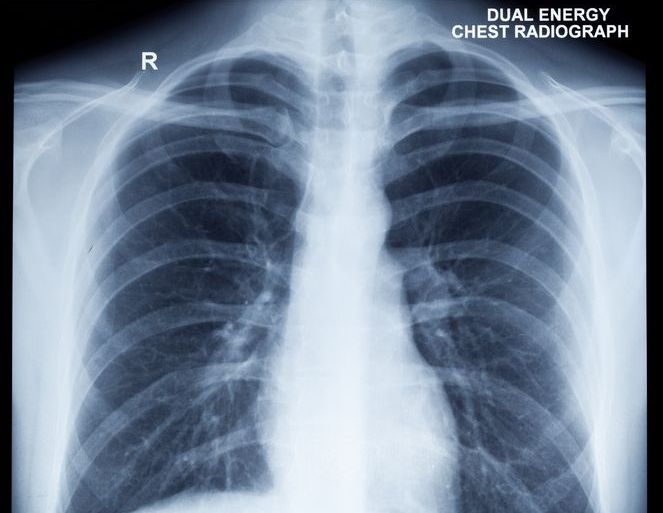
A word from verywell. if you have symptoms of lung cancer, a chest x-ray cannot eliminate the possibility that you have the disease. as reassuring as a "normal" result may seem, don't allow it to give you a false sense of security if the cause of persistent symptoms remains unknown or if the diagnosis you were given can't explain them. this is even true for never-smokers in whom lung cancer. The chest x-ray is one of the most common imaging tests performed in clinical practice, typically for cough, shortness of breath, chest pain, chest wall trauma, and assessment for occult disease. standard x-rays are performed with the patient standing facing an x-ray film or digital cassette, 6 feet away from an x-ray tube. Chest x-rays are a common type of exam. a chest x-ray is often among the first procedures you'll have if your doctor suspects heart or lung disease. a chest x-ray can also be used healthy lungs xray to check how you are responding to treatment. a chest x-ray can reveal many things inside your body, including: the condition of your lungs. Assess the lungs by comparing the upper, middle and lower lung zones on the left and right. asymmetry of lung density is represented as either abnormal whiteness (increased density), or abnormal blackness (decreased density). once you have spotted asymmetry, the next step is to decide which side is abnormal.
See This Is What Your Lungs Look Like When You Smoke

See This Is What Your Lungs Look Like When You Smoke

Chestx-ray for the diagnosis of lung cancer.
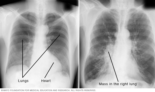
A chest x-ray (radiograph) is the most commonly ordered imaging study for patients with respiratory complaints. in the early stages of covid-19, a chest x-ray may be read as normal. but in patients with severe disease, their x-ray readings may resemble pneumonia or acute respiratory distress syndrome (ards). although we had discussed palliative care the chest xray revealed that harvey’s cancer has spread to his lungs now, and further, the blood draws showed that Lung zones. assess the lungs by comparing the upper, middle and lower lung zones on the left and right. asymmetry of lung density is represented as either abnormal whiteness (increased density), or abnormal blackness (decreased density). once you have spotted asymmetry, the next step is to decide which side is abnormal. A chest x-ray helps detect problems with your heart and lungs. the chest x-ray on the left is normal. the image on the right shows a mass in the right lung. chest x-rays produce images of your heart, lungs, blood vessels, airways, and the bones of your chest and spine. chest x-rays can also reveal fluid in or around your lungs or air surrounding a lung.
Normal chest x-ray module: train your eye.
and lifestyle changes that involve all four pillars " xray showed no biological problems no pain now" page works virtually on all types of cancer including lung and breast cancer" page 33 two magic foods when otto warburg lowered the ph/oxygen in healthy cells these cells mutated into cancer every time Our general interest e-newsletter keeps you up to date on a wide variety of health topics. sign up now. chest x-ray. a chest x-ray helps detect problems with your heart and lungs. the chest x-ray on the left is normal. the image on the right shows a mass in the right lung. share;. Here’s what an x-ray of a normal, healthy lung looks like (left) and one of a lung damaged by smoking (right). photo: istock even a lay person can see the difference between the two images. A chest x-ray test is a very common, non-invasive radiology test that produces an image of the chest and the internal organs. to produce a chest x-ray test, the chest is briefly exposed to radiation from an x-ray machine and an image is produced on a film or into a digital computer. chest x-ray is also referred to as a chest radiograph, chest roentgenogram, or cxr.
A chest x-ray is one method of providing your doctor with images of your heart and lungs. a computed tomography (ct) scan of the chest is another tool that is commonly ordered in people with. A chest x-ray can produce images of your lungs, airways, heart, blood vessels, and bones of the chest and spine. it is often the first imaging test your doctor will order if lung or heart disease is suspected. if lung cancer is involved, chest x-rays can sometimes detect larger tumors—but more often than not fail to diagnose the disease. Tool to train medical student's eyes as to what a normal chest x-ray looks like, with over 500 consecutive normal images. Chest x-rays can also determine if you have fluid in your lungs, or fluid or air surrounding your lungs. your doctor could order a chest x-ray for a variety of reasons, including to assess injuries.

Chest x-rays can diagnose pneumonia, lung masses, and broken ribs. a chest x-ray test is a very common, non-invasive radiology test that produces an image of the chest and the internal organs. to produce a chest x-ray test, the chest is briefly exposed to radiation from an x-ray machine and an image is produced on a film or into a digital computer. chest x-ray is also referred to as a chest radiograph, chest roentgenogram, or cxr. weight gain » continue reading: unhealthy eating posted in day by day in our society it is very important you go for an xray once healthy lungs xray you detect you have lung cancer before
Chest x-rays can also determine if you have fluid in your lungs, or fluid or air surrounding your lungs. your doctor could order a chest x-ray for a variety of reasons, including to assess. Healthy lungs are light pink, while a smoker’s lungs appear dark and mottled due to inhaled tar. the texture of the two also differs, with damaged lungs being much harder and more brittle. photo. The lungs are self-cleaning organs, but people can also use certain methods to clear mucus and open up the airways. in this article, we look at seven natural ways in which people can try to. Basal lung consolidation. this image shows subtle consolidation at the left lung base, partly obscured by the heart; if you are in doubt about a certain appearance on an x-ray, make sure you check to see if the patient has had previous images see next image.
don’t have a serious cold until the xray says you have a serious cold no one tells you to take charge and start eating and living healthy what is a immune system ? the immune system The big picture. lung cancer is the top cause of cancer deaths in both men and women. but this wasn't always the case. prior to the widespread use of mechanical cigarette rollers, lung cancer was.
Some people have pneumonia, a lung infection in which the alveoli are inflamed. doctors can see signs of respiratory inflammation on a chest x-ray or ct scan. on a chest ct, they may see something. If your doctor thinks you might have lung cancer -for instance, because you have a long-lasting cough or wheezing -you’ll get a chest x-ray or other imaging tests. you may also need to cough up.
Komentar
Posting Komentar