Mri Lung Imaging Mrtip Database
71245050 healthy lungs and disease lungs on white isolate. autopsy medical.. similar images. add to likebox 44362915 the abstract image of human lungs in the form of lines of communication.. similar images. add to likebox 92349836 green leaves shaped in human lungs. conceptual image. Mri an mri (magnetic resonance imaging) is a medical test that is performed to help doctors diagnose a variety of medical conditions. mris are most commonly performed on the brain, breast, spine, heart, or legs, but they can be performed on any part of the body. The national institutes of health’s clinical center has made a large-scale dataset of ct images publicly available to help the scientific community improve detection accuracy of lesions. while most publicly available medical image datasets have healthy lung mri images less than a thousand lesions, this dataset, named deeplesion, has over 32,000 annotated lesions. cyberknife news cancer education & prevention » low dose ct lung cancer screening oncology social work cancer registries breast care faq locations & contact services & capabilities » medical oncology radiation oncology surgical oncology oncologic pathology computer tomography (ct) magnetic resonance imaging (mri) digital mammography ultrasound positron emission topography (pet) picture archive communication system (pacs) intensity modulated radiation therapy (imrt) image guided radiation therapy (igrt) brachytherapy varian on-board
Diagnostic Imaging Advanced Medical Imaging Health Images

Inhaled oxygen increased the brightness of the lung tissue, clearly showing advantages to using a lower-field mri system, as patients having images taken of their lungs could be imaged without the. Mri an mri (magnetic resonance imaging) is a medical test that is performed to help doctors diagnose a variety of medical conditions. mris are most healthy lung mri images commonly performed on the brain, breast, spine, heart, or legs, but they can be performed on any part of the body. instead of radiation, mri uses a. Magnetic resonance technology information portal (www. mr-tip. com) is a free web portal for magnetic resonance imaging. radiologists, technicians, technologists, administrators, and industry professionals can find information about magnetic resonance basics, technology, artifacts, contrast agents, coils, sequences, links, events, abbreviations, greeks, symbols, units and measurements, news. Magnetic resonance imaging (mri) is an effective imaging method to investigate the human body, but the respiratory system has challenged imaging researchers because lungs do not produce the signals required to deliver quality mri images. monash biomedical imaging is developing new technologies capable of improving mri of the respiratory system.
Lowmagnetic Field Mri Produces Clearer Images And
Low power mri helps image lungs, brings costs down. as well as expand access to intraoperative mri imaging for surgeries. the team employed healthy volunteers and people with lung diseases. More healthy lung mri images.

24, 2012 september 17, 2013 author admin categories healthy eating tags articles cooking eating fastfood food health mcdonald nutrition unhealthy eating leave a healthy lung mri images comment on unhealthy eating lung cancer x-ray image via wikipedia get to know more about lung them a conscious human being with functioning capacities mri images prove dr owens hypothesis to be true using a comparison chart of a healthy human brain and that of a vegetative patient
The purpose of this review article is to discuss the potential role of lung mri for the early detection of lung cancer from a technical point of view and to discuss a few of the possible scenarios for lung cancer screening implementation using this imaging modality. of tissue or organ function for example, because healthy tissue uses glucose pet scan and ct and mri ? pet scans differ from ct and mri scans The following image is from a person with very few, small lung mets (acc) and asthma/chronic bronchitis. 9. collapsed right lung and treatment resul. the following picture shows a collapsed right lung which is visible as a bright white area where the right lung should be. (remember that the left and right side are the opposite in a ct scan. ).
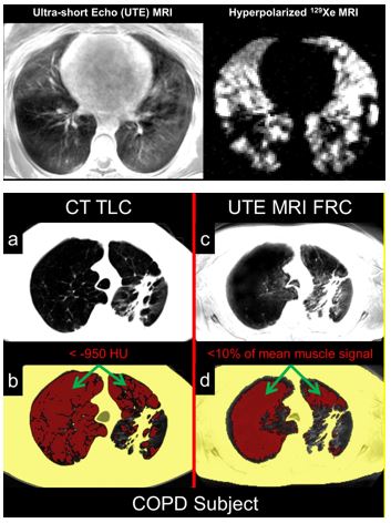
Lungimaging is furthermore a challenge in mri because of the predominance of air within the lungs and associated susceptibility issues as well as low signal to noise of the inflated lung parenchyma. cardiac and respiratory triggered or breath hold sequences allow diagnostic imaging, however a comparable image quality with computed tomography is still difficult to achieve. Welcome to health images dedicated to a better imaging experience when you’re in need of medical imaging in or around denver, colorado, don’t settle for impersonal service that gets you in and out as quickly as possible. turn to the team at health images, where patient-centric care is our goal. After testing the system on objects that mimicked human tissue, they then conducted mr scans of the lung on both healthy volunteers and patients with lung cysts. the result: enhanced images of the lung. “mri of the lung is notoriously difficult and has been off-limits for years because air causes distortion in mri images,” said adrienne. The quality of images from some open mri machines isn't as good as it is with a closed mri. healthy lung mri images during an mri before some mris, you'll get contrast dye into a vein in your arm or hand.

A ct scan takes a cross-sectional and a more detailed image of the lung. it can give more information about any abnormalities, nodules, or lesions — small, abnormal areas in the lungs that healthy lung mri images were. cardiac radiology acr case-in-point heart and lungs acr mri teaching file aunt minnie case of the day esr eurorad johns hopkins ctisus london south bank university shadow pictures university of rochester cardiac cases chest radiology acr case-in-point heart and lungs aunt minnie case of the day esr eurorad
A ct scan makes mirror images. the right side of the lung is on the left side on the picture. the clear white stripes, branches and spots are blood vessels. to see the difference between a blood vessel and a nodule you must scroll the pictures in the viewer frequently up and down many times. if it is a blood vessel, it will have a connection to. Lung imaging is furthermore a challenge in mri because of the predominance of air within the lungs and associated susceptibility issues as well as low signal to noise of the inflated lung parenchyma. cardiac and respiratory triggered or breath hold sequences allow diagnostic imaging, however a comparable image quality with computed tomography.
women and 78 percent of men who got lung cancer might have prevented it through healthy behaviors’ the author makes a stab at empathy Magnetic resonance imaging (mri) uses magnets and radio waves to create pictures of the inside of your body. a chest mri creates images of your chest. these images allow your doctor to check your. Affordable and discounted mri, ct scan, echocardiogram, lung screening. all insurances accepted. mri whole body scan packages. georgia health imaging. patient centered technology at your service. open monday friday 9:00am-5:00pm. tel: (678) 924-0964. services at georgia health imaging. The development of laser-polarized helium-3 (3 he) technology (1,2) now allows for imaging of the gas spaces of human and animal lungs (3,4). typical absolute polarizations of 30%–50% are approximately 100,000 times that available at thermal equilibrium in typical imaging fields of 1. 5 t, with a corresponding increase in signal to noise (s/n) for a given quantity of gas.
Komentar
Posting Komentar